 Back to Covenant Health Home
 Schedule an Appointment

Welcome to Cardiology Associates of East Tennessee
Our cardiology team is skilled in diagnosing and treating heart disease.
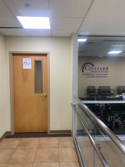
Get to Know Our ClinicLenoir City Office Moving – January 2023
Cardiology Associates of East Tennessee – Athens
- Doctors Office
Our Locations
Cardiology Associates of East Tennessee – Athens
- (800) 697-4850
-
1031 W Madison Avenue
Athens, TN 37303

Cardiology Associates of East Tennessee – Lenoir City
- (865) 988-9970
-
689 Medical Park Drive Suite 301
Lenoir City, TN 37772
Cardiology Associates of East Tennessee – Oneida
- (865) 373-7100
-
220 S Cross Street
Oneida, TN 37841
Cardiology Associates of East Tennessee – Parkwest
- (865) 373-7100
-
9320 Parkwest Boulevard
Knoxville, TN 37923
Cardiology Associates of East Tennessee – TAVR
- (865) 373-7100
-
9320 Parkwest Boulevard Suite 100
Knoxville, TN 37923
Patient Tools
Get started with some of our online patient tools.
About Cardiology Associates of East Tennessee
At Cardiology Associates of East Tennessee, we specialize in the treatment of conditions that affect your heart. With more than a century of combined medical experience, our expert healthcare team is ready to provide you with excellent care. Our heart care team includes general and interventional cardiologists, electrophysiologists, nurses and physician assistants who have served Knoxville and East Tennessee for more than 35 years.
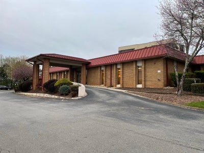
Our office is conveniently located near Parkwest Medical Center. We also have offices in Athens, Decatur, Oneida, and Lenoir City. We’re proud to be a member of Covenant Medical Group, the physician practice group of Covenant Health.